
About Conference
Plastic Surgery 2027
Plastic Surgery 2027 – 2nd International Conference on Plastic Surgery, Aesthetics & Skin Rejuvenation will be held on July 19–21, 2027, in Vienna, Austria, bringing together leading plastic surgeons, aesthetic specialists, dermatologists, cosmetic physicians, researchers, clinicians, delegates, and industry professionals from around the world. Guided by the theme “Innovating Aesthetic Medicine and Plastic Surgery for Natural, Safe and Future-Ready Outcomes,” the conference provides a dynamic global platform featuring renowned keynote speakers, scientific sessions, oral and poster presentations, panel discussions, and industry exhibitions focused on the latest advancements in plastic surgery, aesthetic medicine, and skin rejuvenation. The event fosters strong networking and collaboration opportunities by connecting academia, healthcare institutions, aesthetic clinics, and medical technology companies, enabling knowledge exchange, research partnerships, and professional growth. Plastic Surgery 2027 highlights...
Conference Starts In:
Plastic Surgery, Aesthetics & Skin Rejuvenation
Scientific Sessions
Explore innovative scientific sessions in Plastic Surgery, Aesthetics & Skin Rejuvenation
- Regulatory Standards and Legal Aspects of Aesthetic Surgery
- Combination Therapies in Modern Aesthetic Practice
- Evidence-Based Approaches in Cosmetic and Plastic Surgery
- Cultural Perspectives and Patient Expectations in Aesthetics
- Practice Management and Business Models in Aesthetic Surgery
- Emerging Trends in Aesthetic and Plastic Surgery
- Postoperative Care and Optimizing Surgical Outcomes
- Hair Restoration and Scalp Rejuvenation Techniques
- Cosmetic Dermatology and Advanced Skin Treatment Modalities
- Patient Safety, Ethics, and Risk Management in Surgery
- Artificial Intelligence Applications in Aesthetic Medicine
- Digital Planning and 3D Imaging in Plastic Surgery
Committee Members
Our organizaing committee members
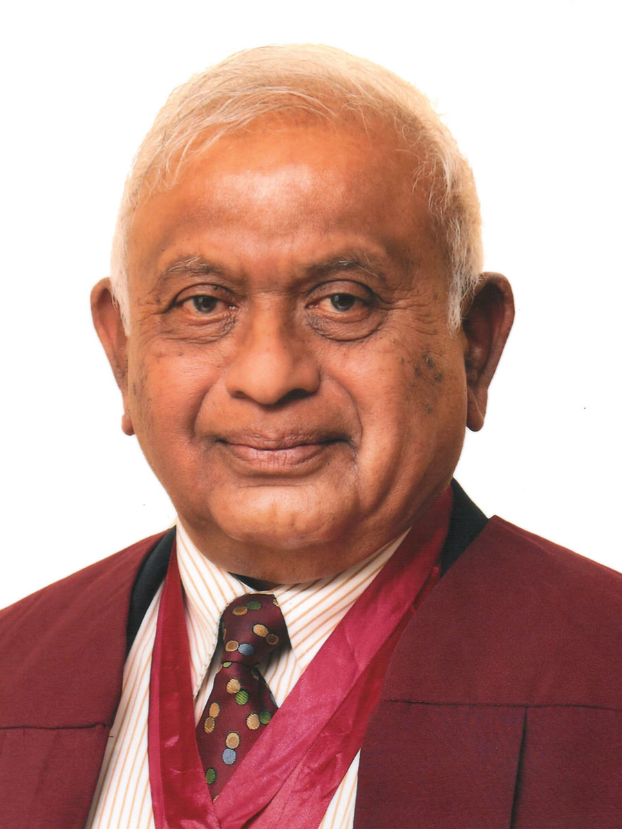
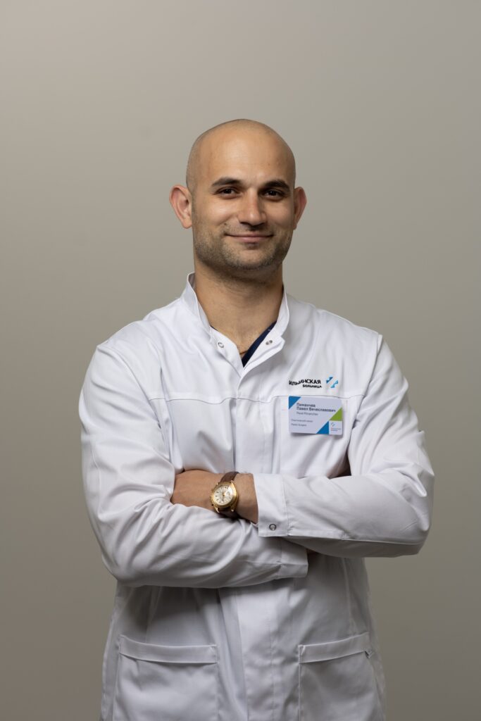 Russia
Pavel Vyacheslavovich Pimanchev
Committee Member
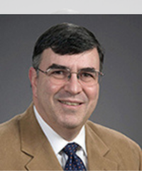 USA
USA
Jeffrey E. Rubenstein
Committee Member
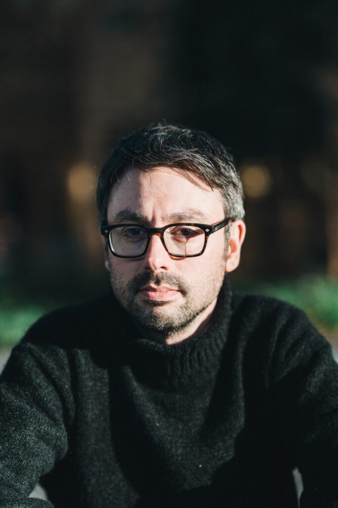 USA
USA
Francesco
Committee Member
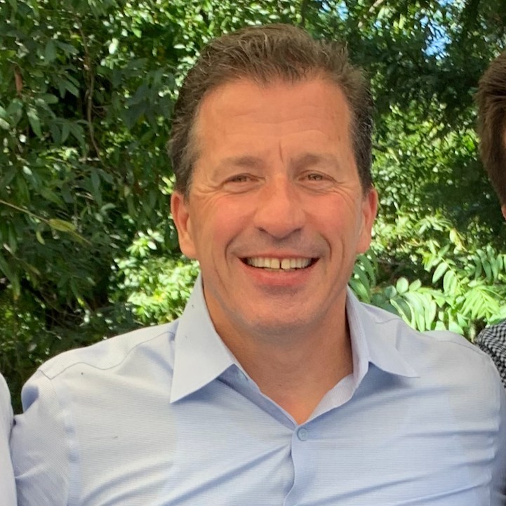 Italy
Italy
Giovanni Leoni
Committee Member
Hotel Services & Amenities
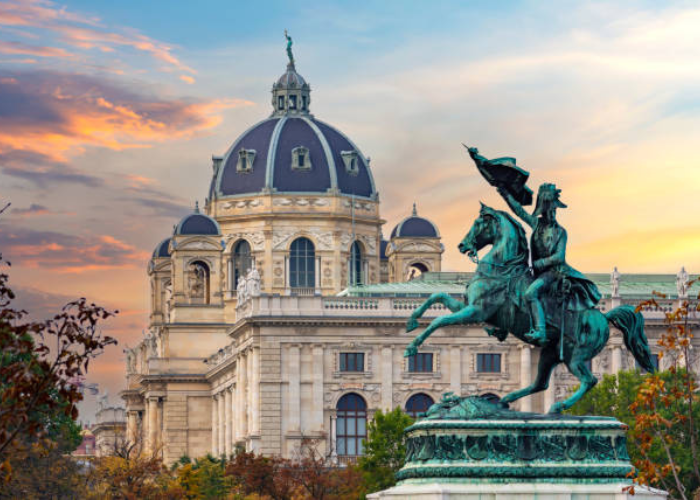
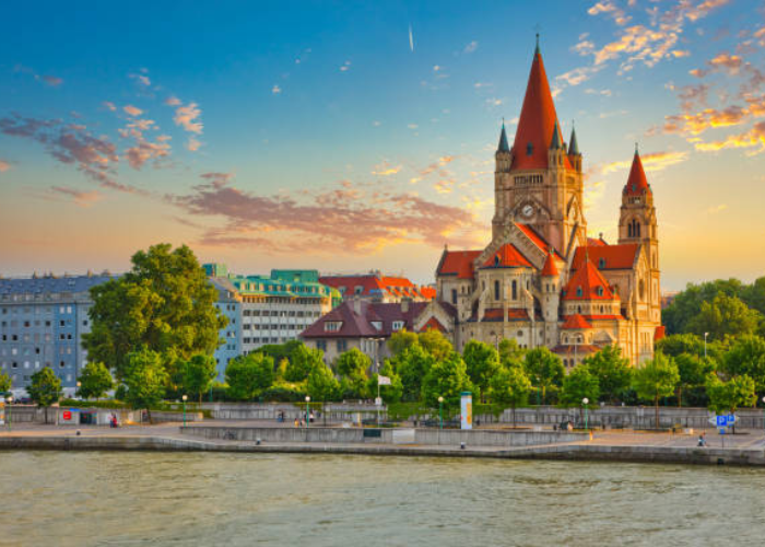
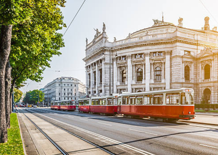
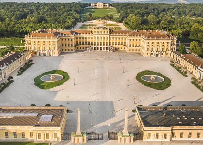
Conference Venue DetailsVienna, Austria, is a world renowned city known for its excellence in healthcare, education, research, and innovation. It is an ideal destination for international scientific and medical conferences. During July, the city enjoys a pleasant summer climate with comfortable temperatures. Vienna is home to leading universities, research institutions, and major medical centers that support academic collaboration. The city offers famous attractions such as Schönbrunn Palace, St. Stephen’s Cathedral, and the Hofburg Palace. Delegates can enjoy rich cultural experiences, historic neighborhoods, classical music, and Austrian cuisine. Vienna is easily accessible through Vienna International Airport with global flight connections. Local transportation includes an efficient metro, trams, buses, and taxis. The city offers modern conference facilities and reliable infrastructure. Attending a conference in Vienna provides both professional growth and a memorable travel experience.
-
Audio/Visual Equipment Rental
-
High-Speed Wireless Internet
-
Dedicated On-site Parking
-
Guest Room Accommodation Packages
Registration Offers
Our registration offers
Speaker Registration
Global Stage for Your Research
- Global Recognition
- Abstract Publication
- Certificate of Honor
- Networking Access
Student Registration
Empowering the Next Generation
- Expert Mentorship
- Career Development
- Reduced Pricing
- Participation Award
Delegate Registration
Immersive Learning & Networking
- Knowledge Exchange
- Premium Materials
- Interactive Learning
- All-Inclusive Catering
Global Perspectives, Shared Success
Discover how our summits have empowered researchers and industry leaders to shape the future of their fields.
Read MoreThank you for the beautiful photos; Thank you for the conference invitation. The event was truly inspiring and meaningful. They are a wonderful reminder of the significant event... Read more

Thanks so much for your kind words! I had a great experience at the conference. As I mention in the video, I got to share my work with some truly brilliant speakers and... Read more
I am grateful for the invitation to the traditional medicine, immunology, biochemistry and food technology event. I consider that the event met my expectations of meeting other... Read more
Thank you for organizing a great event, it was a good experience.
I returned home after a fruitful conference. I had the opportunity to upgrade my knowledge and interact with many active scientists. My presentation was well received. Thank you... Read more
Thank you for organizing the Microbiology conference; it was a successful event that discussed the current issues of microbiology science in medicine, food, and other fields.

Thank you very much for the organization. It was really good conference and we enjoyed a lot those moments. I will try to send you a short video about my impressions next week but... Read more
The conference is very nice.
Thank you for your kind words and for organizing the International Conference on Immunology & Vaccination. It was a pleasure to participate and contribute to the discussions.... Read more

I hope this email finds you well. Thank you very much for having me. I have finished the conference and returned to China safely. I had a wonderful week in Zurich and thank you... Read more

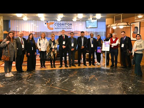
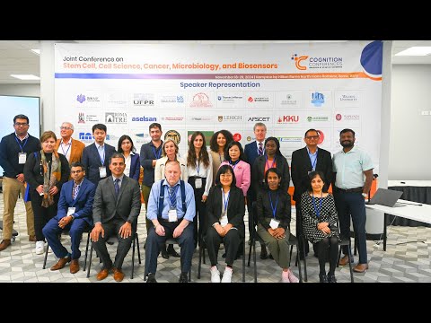
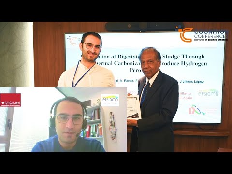
Video Gallery
Watch highlights, speaker insights, and previews of upcoming events.
Official Media Partners
We are proud to collaborate with leading media organizations.


Get In Touch
To know more about our conferences and events do get in touch with us and join our large network of scientists, professional experts, and research scholars.
Contact Information
-
Office Address ProCognition Conferences OÜ
Kaupmehe tn 7-120, Kesklinna district
Tallinn city, Harju county 10114
Estonia
VAT: EE102850103 -
WhatsApp Number +372 668 2606
-
E-mail address contact@cognitionconferences.org